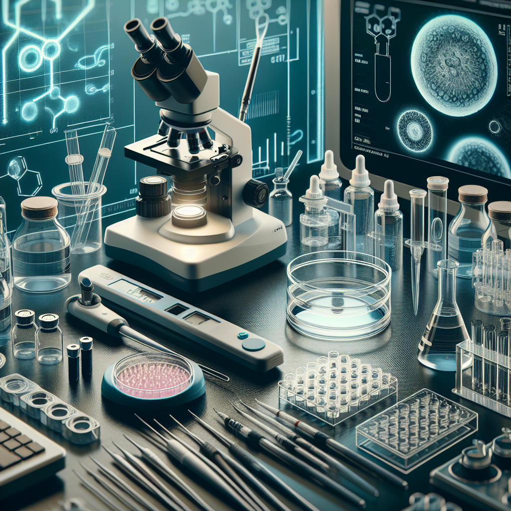
Assisted Reproductive Technology (ART) helps individuals and couples overcome infertility. ART procedures rely on specialized equipment to increase the chances of successful conception. This blog explores the key equipment used in ART, their functions, and how they work.
1. Ultrasound Machines
Ultrasound machines use high-frequency sound waves to create images of internal organs. In ART, they focus on visualizing the ovaries, uterus, and developing follicles. These images help doctors track ovulation, monitor follicular growth, and guide embryo transfers. It uses sound waves, not radiation, making it safe for the person undergoing ART or the developing fetus.
Working Principle of Ultrasound Scans
Ultrasound imaging relies on the transmission and reflection of high-frequency sound waves. Here’s how it works:
- Sound Wave Emission: The ultrasound probe (transducer) emits high-frequency sound waves into the body.
- Wave Interaction: These sound waves travel through tissues and bounce back when they encounter different structures, such as organs or fluids.
- Echo Reception: The transducer detects the reflected sound waves (echoes).
- Image Formation: The machine processes these echoes to create real-time images on a monitor.
The difference in the speed and intensity of the reflected sound waves helps the machine differentiate between various tissues, creating a detailed image of the internal structures.
How Ultrasound Machines Work
Transvaginal Ultrasound
- Probe Insertion: Insert a slender probe into the vagina.
- Image Capture: The probe emits sound waves that bounce off the ovaries and follicles.
- Real-Time Images: The machine converts these waves into real-time images on a screen.
Abdominal Ultrasound
- Transducer Application: Apply a gel on the abdomen and move the transducer over it.
- Sound Wave Emission: The transducer emits sound waves that penetrate the abdominal wall.
- Image Display: The machine translates these waves into images of the uterus and follicles.
Doppler Ultrasound
- Blood Flow Measurement: The Doppler ultrasound measures blood flow in the reproductive organs.
- Color-Coded Images: The machine produces color-coded images to show blood flow patterns.
Importance of Ultrasound Machines in ART
Follicular Monitoring
- Track Growth: Monitor the growth of ovarian follicles.
- Timing: Determine the best time for ovulation induction and egg retrieval.
- Adjustments: Adjust medication dosages based on follicular development.
Ovulation Induction
- Identify Maturity: Ensure follicles reach the right size before triggering ovulation.
- Schedule Retrieval: Plan the timing for egg retrieval accurately
Embryo Transfer
- Placement: Guide the catheter to place embryos precisely in the uterus.
- Check Position: Ensure optimal positioning of the embryos for implantation.
Pregnancy Monitoring
- Early Detection: Confirm early pregnancy and check for multiple gestations.
- Fetal Health: Monitor the health and development of the fetus.
The Purpose of Applying Gel in Ultrasound Scans
During an ultrasound scan, applying gel on the skin serves several critical purposes. This seemingly simple step significantly enhances the quality and accuracy of the ultrasound images. Ultrasound gel is a specialized, water-based formulation designed to optimize the performance of ultrasound imaging. Its primary components include water, glycerin or propylene glycol, thickening agents, and preservatives. Together, these ingredients ensure that the gel remains effective as a medium for sound wave transmission, providing clear and accurate images while ensuring patient comfort and safety.
Purpose of the Gel
- Transmission Medium: The gel acts as a conductive medium that allows ultrasound waves to pass efficiently from the transducer into the body.
- Eliminates Air Pockets: By filling in any small air gaps between the transducer and the skin, the gel ensures continuous contact and effective wave transmission.
- Lubrication: The gel provides a slippery surface, allowing the transducer to glide smoothly over the skin, which enhances the comfort of the procedure for the patient.
2. Incubators
Incubators provide a controlled environment that mimics the natural conditions of the uterus, ensuring optimal embryo development. By maintaining precise temperature, humidity, and gas levels, incubators increase the chances of successful ART outcomes. Their role in creating a stable and sterile environment is indispensable, supporting various stages of embryo development and ultimately contributing to higher success rates in ART procedures. As technology advances, incubators continue to improve, offering even better conditions and monitoring capabilities for embryo growth.
How Incubators Work
Temperature Control
- Consistent Heat: Incubators maintain a constant temperature, usually around 37°C (98.6°F), similar to the human body.
- Heating Elements: They use heating elements to provide and regulate this temperature.
- Temperature Sensors: Sensors continuously monitor the temperature to prevent fluctuations.
Humidity Control
- Maintaining Moisture: Incubators keep the humidity levels around 95%. This prevents the embryos from drying out.
- Water Reservoirs: They include water reservoirs to maintain high humidity levels.
- Humidity Sensors: Sensors ensure the humidity stays within the desired range.
Gas Concentration
- CO2 Levels: Incubators maintain a CO2 concentration of about 5-6% to keep the pH levels of the culture media stable.
- O2 Levels: They often reduce oxygen levels to around 5%, mimicking the lower oxygen concentration in the human reproductive tract.
- Gas Sensors: These sensors monitor and adjust the CO2 and O2 levels as needed.
Monitoring Systems
- Continuous Monitoring: Advanced incubators continuously monitor environmental conditions.
- Alarm Systems: They have alarm systems to alert staff if conditions go out of range.
- Data Logging: Some incubators log data for future reference and analysis.
Importance of Incubators in ART
Optimal Embryo Development
- Stable Environment: Incubators provide a stable environment essential for embryo growth.
- Mimicking Natural Conditions: By mimicking the uterus, they ensure embryos develop as they would inside the body.
Increased Success Rates
- Higher Viability: Maintaining optimal conditions increases the chances of embryo viability.
- Better Outcomes: This leads to higher success rates in ART procedures.
Reduced Contamination Risk
- Sterile Environment: Incubators provide a sterile environment, reducing the risk of contamination.
- Controlled Access: Many have features like controlled access to minimize exposure to external contaminants.
Supporting Various Stages
- Fertilization: Incubators support the initial stages of fertilization.
- Early Development: They are crucial during the early stages of embryo development, from zygote to blastocyst.
Types of Incubators
Benchtop Incubators
- Compact Size: These are small and fit on laboratory benches.
- Easy Access: They offer easy access for routine checks and procedures.
Larger Incubators
- Higher Capacity: These can hold more samples at once.
- Advanced Features: They often come with more advanced monitoring and control features.
Time-Lapse Incubators
- Continuous Observation: These incubators have built-in cameras to continuously monitor embryo development.
- Detailed Analysis: They provide detailed images and videos, helping embryologists track development without disturbing the embryos.
3. Microscopes
Microscopes magnify small objects, making them visible and easier to analyze. In ART, they provide detailed views of sperm, eggs, and embryos. This magnification helps embryologists assess the quality and development of these cells, guiding crucial steps in the reproductive process.
How Microscopes Work
Basic Principle
- Magnification: Lenses within the microscope magnify the object being observed.
- Light Source: A built-in light source illuminates the object, enhancing visibility.
- Resolution: High-resolution optics provide clear and detailed images.
Types of Microscopes Used in ART
- Light Microscopes:
- Standard Use: Commonly used for routine examinations of sperm, eggs, and embryos.
- Functionality: Utilize visible light to illuminate and magnify the specimen.
- Stereomicroscopes:
- Three-Dimensional View: Provide a three-dimensional view of the specimen.
- Manipulation: Ideal for handling and manipulating embryos during procedures like Intracytoplasmic Sperm Injection (ICSI).
- Inverted Microscopes:
- Cell Culture: Specifically designed for observing cells in culture dishes.
- Bottom Illumination: Light source is located below the stage, allowing observation from underneath.
- Phase-Contrast Microscopes:
- Enhanced Contrast: Enhance contrast in transparent specimens without staining.
- Live Cells: Useful for observing live cells and embryos.
- Electron Microscopes:
- High Magnification: Provide much higher magnification than light microscopes.
- Detailed Structure: Used for detailed study of sperm morphology and other cellular structures.
Specialized Techniques
- Intracytoplasmic Sperm Injection (ICSI):
- Micromanipulators: Attach micromanipulators to the microscope for precise handling of sperm and eggs.
- Single Sperm Injection: Inject a single sperm into an egg under high magnification.
- Preimplantation Genetic Diagnosis (PGD):
- Cell Analysis: Use microscopes to biopsy and analyze cells from embryos.
- Genetic Screening: Examine embryos for genetic abnormalities before implantation.
Importance of Microscopes in ART
Sperm Analysis
- Motility Assessment: Evaluate sperm motility and movement.
- Morphology Examination: Assess the shape and structure of sperm.
- Concentration Measurement: Determine sperm concentration in a sample.
Egg Quality Assessment
- Maturity Check: Examine eggs to ensure they are mature enough for fertilization.
- Structural Integrity: Check for abnormalities or defects in the eggs.
Embryo Evaluation
- Development Monitoring: Observe and assess embryo development stages.
- Grading: Grade embryos based on quality and development, guiding selection for transfer.
Precision in Procedures
- ICSI Accuracy: Ensure precise sperm injection into the egg during ICSI.
- Embryo Biopsy: Perform accurate biopsies for genetic testing and screening.
Enhancing Success Rates
- Informed Decisions: Provide detailed information to make informed decisions about embryo selection.
- Reduced Risks: Minimize risks by selecting the best quality sperm, eggs, and embryos.
4. Cryopreservation Equipment
Cryopreservation involves freezing biological material at extremely low temperatures to preserve its viability. In ART, this means storing sperm, eggs, and embryos for extended periods without compromising their quality. The process relies on cryoprotectants to prevent ice crystal formation, which can damage cells.
How Cryopreservation Equipment Works
Freezing Process
- Cryoprotectant Addition:
- Add cryoprotectants to the biological material to protect cells from damage during freezing.
- Common cryoprotectants include dimethyl sulfoxide (DMSO) and glycerol.
- Cooling Rate:
- Use controlled-rate freezers to gradually lower the temperature.
- Slow cooling allows cells to adapt to the cold and prevents ice crystal formation.
- Vitrification:
- Employ vitrification for rapid freezing, turning the material into a glass-like state.
- This method uses high concentrations of cryoprotectants and ultra-rapid cooling.
Storage
- Liquid Nitrogen Tanks:
- Store frozen samples in liquid nitrogen tanks at temperatures around -196°C (-320.8°F).
- Ensure the tanks maintain a stable temperature to preserve sample viability.
- Storage Containers:
- Use specialized storage containers, such as cryovials and straws, to hold the samples.
- Label containers with detailed information for easy identification and tracking.
Thawing Process
- Rapid Warming:
- Thaw frozen samples quickly to minimize ice crystal formation during warming.
- Use a warm water bath or incubator to bring samples to the appropriate temperature.
- Cryoprotectant Removal:
- Gradually remove cryoprotectants to prevent osmotic shock to the cells.
- Use a stepwise dilution process to safely return cells to their natural state.
Importance of Cryopreservation Equipment in ART
Fertility Preservation
- Cancer Patients:
- Preserve fertility for individuals undergoing cancer treatment that may affect reproductive health.
- Freeze sperm, eggs, or embryos before starting chemotherapy or radiation.
- Delayed Childbearing:
- Allow individuals or couples to delay childbearing for personal or medical reasons.
- Store gametes or embryos for future use.
ART Flexibility
- Multiple Cycles:
- Use stored embryos in multiple ART cycles, reducing the need for repeated egg retrievals.
- Increase the chances of successful pregnancy over several attempts.
- Embryo Selection:
- Freeze surplus embryos during an ART cycle for future use.
- Select the best quality embryos for transfer after initial cycles.
Genetic Testing
- Preimplantation Genetic Diagnosis (PGD):
- Freeze embryos after biopsy for genetic testing.
- Transfer only healthy embryos, reducing the risk of genetic disorders.
Donor Programs
- Egg and Sperm Donation:
- Preserve donor eggs and sperm for use in donor programs.
- Offer a larger pool of donor options for individuals and couples.
Increased Success Rates
- Optimal Timing:
- Schedule embryo transfers at the most optimal time for the recipient.
- Align with the recipient’s natural or prepared uterine environment.
- Reduced Stress:
- Allow time for physical and emotional recovery between cycles.
- Reduce the stress associated with fresh cycles and immediate transfers.
For further insights on cryogenic technology, check out the article Cryogenic Technology: The Cold Frontier Transforming Our Lives
5. Egg Retrieval Needles
Egg retrieval needles are specialized medical instruments designed to extract eggs from the ovarian follicles. This process, known as follicular aspiration, involves using ultrasound guidance to accurately locate and retrieve the eggs. The procedure requires precision and care to maximize egg recovery and minimize patient discomfort.
How Egg Retrieval Needles Work
Preparation
- Ovarian Stimulation:
- Administer hormonal medications to stimulate the ovaries.
- This encourages the development of multiple follicles containing mature eggs.
- Monitoring:
- Use ultrasound and blood tests to monitor follicle growth and hormone levels.
- Schedule the egg retrieval procedure when follicles reach the optimal size.
The Procedure
- Ultrasound Guidance:
- Use a transvaginal ultrasound probe to visualize the ovaries and follicles.
- Guide the egg retrieval needle to the target follicles accurately.
- Needle Insertion:
- Insert the needle through the vaginal wall and into the ovarian follicles.
- The needle is long, thin, and hollow, designed for precise aspiration.
- Follicular Aspiration:
- Apply gentle suction to the needle to aspirate the follicular fluid.
- Collect the fluid, which contains the mature eggs, into a test tube.
- Egg Collection:
- Transfer the aspirated fluid to the embryology lab.
- Embryologists identify and isolate the eggs from the fluid under a microscope.
Needle Design
- Sharp Tip:
- The needle tip is sharp to penetrate the follicle wall smoothly.
- Minimizes tissue trauma and discomfort for the patient.
- Echo-Reflective Coating:
- Some needles have an echo-reflective coating to enhance visibility on ultrasound.
- This coating helps guide the needle precisely to the follicles.
- Flexible Shaft:
- The needle shaft is flexible, allowing for better maneuverability.
- Facilitates access to follicles from different angles.
Importance of Egg Retrieval Needles in ART
Maximizing Egg Recovery
- Precision:
- Accurate needle placement ensures maximum egg recovery from each follicle.
- High egg yield increases the chances of successful fertilization and embryo development.
- Minimizing Damage:
- The design and sharpness of the needle reduce tissue damage and bleeding.
- Preserves ovarian function and patient comfort.
Enhancing Success Rates
- Multiple Eggs:
- Retrieving multiple eggs increases the likelihood of obtaining viable embryos.
- Provides more options for embryo selection and transfer.
- Cryopreservation:
- Extra eggs can be frozen for future use.
- Offers flexibility for additional ART cycles without repeated retrievals.
Patient Safety and Comfort
- Anesthesia:
- Administer local or general anesthesia or sedation to minimize discomfort during the procedure.
- Ensures a safer and more comfortable experience for the patient.
- Quick Procedure:
- The egg retrieval process typically takes 30-45 minutes, however the procedure time varies with number of follicles developed.
- Patients can usually go home the same day.
6. Embryo Transfer Catheters
Catheters place embryos into the uterus.
Working
- Flexible Design: The catheter’s flexible and thin design minimizes trauma to the uterine lining.
- Guided Transfer: Perform the procedure under ultrasound guidance to ensure accurate placement.
Embryo transfer catheters are vital for the final step in the ART process, ensuring safe and precise embryo transfer.
7. Laboratory Culture Media
Concept and Use
Culture media provide nutrients and an environment for the growth and development of gametes and embryos in vitro.
Working
- Nutrient-Rich Solutions: Contain essential amino acids, vitamins, and minerals that support cellular development.
- Specialized Media: Different stages of ART require specific media formulations.
The quality of culture media significantly impacts the success of ART procedures.
Conclusion
Success in Assisted Reproductive Technology depends on the precise and effective use of various specialized equipment. Each piece, from ultrasound machines to embryo transfer catheters, plays a critical role at different stages of the ART process. By maintaining optimal conditions and allowing meticulous handling of gametes and embryos, this equipment ensures the highest chances of successful conception and healthy pregnancy outcomes. As technology advances, the efficacy and accessibility of ART procedures will likely improve.
